Gallbladder Polyps: Do They Need Surgery?
Need expert consultation? Book an appointment with Dr. Babu Elangovan.
Book AppointmentA polyp on an ultrasound report is one of the more anxiety-producing incidental findings I review in clinic. The word sounds like cancer. Most of the time it is not — but some polyps do need the gallbladder out, and the difference lives in size, shape, growth and context, not in how worried the report makes you feel.
Silent-versus-symptomatic stones are covered in do all gallstones need surgery. Recovery after removal lives in life after gallbladder removal. This article owns polyps — what they are, how we risk-stratify them, and when laparoscopic cholecystectomy is the honest next step.
Polyp vs stone — get this right first
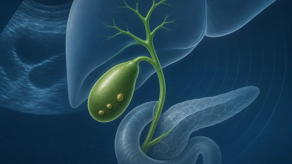
A gallbladder polyp is a growth projecting from the inner wall into the lumen. A gallstone is a mobile deposit of crystallised bile. On ultrasound, stones usually move when you roll and cast an acoustic shadow; polyps stay put and typically do not shadow. Both can sit in the same organ. They are managed by different rules.
| Feature | Polyp | Gallstone |
|---|---|---|
| Nature | Fixed tissue / lipid projection | Mobile crystal |
| Ultrasound | Anchored; usually no shadow | Moves; often shadows |
| Main driver of surgery | Size, growth, neoplastic risk | Symptoms and complications |
How most polyps are found
Most are silent. They turn up on scans done for check-ups, pregnancy, or unrelated abdominal discomfort. Transabdominal ultrasound measures diameter, notes pedunculated (stalk) versus sessile (broad-based) shape, and looks for multiplicity. Sessile morphology generally worries clinicians more than a tiny stalked cholesterol polyp.
Borderline or suspicious cases may need endoscopic ultrasound (EUS), CT or MRCP — not because every polyp needs a scan cascade, but because ultrasound alone cannot always separate a harmless cholesterol deposit from a true adenoma.
Types — cholesterol vs true polyps
Roughly most “polyps” on everyday scans are cholesterol pseudopolyps — lipid-laden macrophages in the mucosa, often multiple and small, with no malignant potential. Inflammatory polyps and adenomyomatosis are also benign patterns.
Adenomas are true epithelial polyps. They are uncommon but matter because they can progress through dysplasia toward gallbladder adenocarcinoma. Imaging cannot reliably label “adenoma” versus “cholesterol” in every case — which is why size and growth thresholds exist instead of waiting for a perfect preoperative biopsy. You cannot safely snare a gallbladder polyp the way we snare a colon polyp; the wall is too thin.
The size and risk ladder (high-level)
Guidelines cluster decisions around millimetres — useful anchors, not a DIY operating licence:
| Size (approx.) | Usual approach |
|---|---|
| Under ~6 mm | Often surveillance; high chance of benign pseudopolyp |
| ~6–9 mm | Closer follow-up; operate sooner if growth, sessile shape or other risk factors |
| ≥ ~10 mm | Surgical evaluation recommended — malignancy risk rises meaningfully |
Growth of about 2 mm or more on serial scans, age and other comorbidities, primary sclerosing cholangitis, concurrent stones, and sessile morphology all push the conversation toward removal even below the classic 10 mm line. Your surgeon integrates the whole picture; a single number on a report is not a verdict.
When surgery is recommended
Cholecystectomy is the definitive treatment when intervention is indicated — usually laparoscopic, sometimes robotic. Indications we discuss most often:
- Polyp ≥ about 10 mm
- Documented growth on surveillance
- Symptoms attributable to biliary disease (even with a smaller polyp)
- High-risk morphology or clinical context (sessile lesion, selected patients with stones or PSC)
The specimen goes to pathology so we finally know what the “polyp” was. Technique and hospital pathway overlap with stone surgery — see gallbladder stone surgery in Chennai and laparoscopic GI surgery — but the indication here is neoplastic risk and growth, not only biliary colic.
Why we remove the whole gallbladder
Patients reasonably ask whether we can “just take the polyp out.” Colon polyps can be snared because the colon wall is thick enough and accessible endoscopically. The gallbladder wall is millimetres thin and sits against the liver bed; attempting local excision risks bile leak, bleeding and incomplete clearance. Removing the organ is therefore the oncologically honest and surgically safe choice when intervention is needed.
Pathology of the opened gallbladder also settles arguments ultrasound cannot: cholesterolosis versus adenoma versus, rarely, early cancer. That answer changes follow-up intensity even when the operation felt “routine.”
What surveillance looks like in practice
If we agree to watch, the plan is not “come back if you worry.” It is a dated repeat ultrasound, preferably at the same centre or with prior images available for side-by-side comparison. Growth is easy to miss when each report is read in isolation. Bring the DICOM or printed prior films — typed millimetres without images are a weak foundation for a surgical decision.
Red flags — do not wait for the next annual scan
Seek prompt review for persistent right-upper-quadrant pain, recurrent vomiting after fatty meals, jaundice, dark urine with pale stools, fever with biliary pain, or unexplained weight loss. Those symptoms can mean stones, inflammation, obstruction or — rarely — advanced disease. Self-flushes and “gallbladder cleanses” have no role and can delay care. Related ductal problems are covered in bile duct stones.
Pain that comes in waves after heavy meals and settles in hours is classic biliary colic — whether driven by stones, sludge, or a gallbladder that is inflamed around a polyp. Fever with continuous pain is not “gas”; it can be cholecystitis and needs same-day assessment. Yellow eyes with dark urine mean bile is not reaching the gut the way it should — imaging that night beats another week of WhatsApp photo advice.
What surgery changes — and what it does not
Laparoscopic cholecystectomy usually means a short stay, a few days of soreness, and a temporary low-fat, smaller-meal phase while bile flows continuously from liver to intestine. Most people return to a normal diet. Detailed recovery belongs to the life after removal guide — not repeated here.
What surgery does achieve for polyps is removing the organ that harbours a lesion we cannot safely sample in situ, and giving pathology a chance to clear or escalate concern.
When to see a GI / HPB surgeon in Chennai
Bring the ultrasound images (not only the typed report), prior scans for growth comparison, and a symptom timeline. We will place you on the watch-versus-operate map rather than defaulting to either panic or indefinite neglect. If a previous doctor already recommended removal and you want a second look at the images before listing for surgery, that is a reasonable use of a specialist visit — especially when the polyp sits near the 10 mm line or reports disagree on size.
Book an appointment or call +91 94440 43478.
This article is general information, not personal medical advice. Size thresholds summarise common clinical practice and must be applied by your surgeon to your scan and history. Seek urgent care for jaundice with fever, severe abdominal pain, or vomiting blood.
Authored by